
Using CT imaging, USC researchers Summer Decker (left) and Jonathan Ford reveal the hidden anatomy of two ancient mummies, uncovering details about health, injury and mummification practices. In the photo, they hold a printed 3D model from CT imaging. (USC Photo/Sean Dube)
Scanning the past: USC researchers unlock ancient secrets with cutting-edge technologies
USC researchers’ CT scans of two Egyptian mummies demonstrate the power of advanced imaging technologies to bridge the gap between the present day and the ancient world.
When Nes-Hor and Nes-Min, two ancient Egyptian mummies, arrived at Keck Hospital of USC on the morning of Jan. 17, Summer Decker was determined to uncover as much detail as possible beneath their linen shrouds.
Decker — the Grace Whisler Professor of Medicine and professor of clinical radiology at the Keck School of Medicine of USC — chose the hospital’s most advanced CT scanner, which captures 320 cross-sectional image “slices” per rotation, for the job. To ensure the highest resolution, she programmed the slices to be as thin as the machine would allow: half a millimeter, the width of a grain of sand.
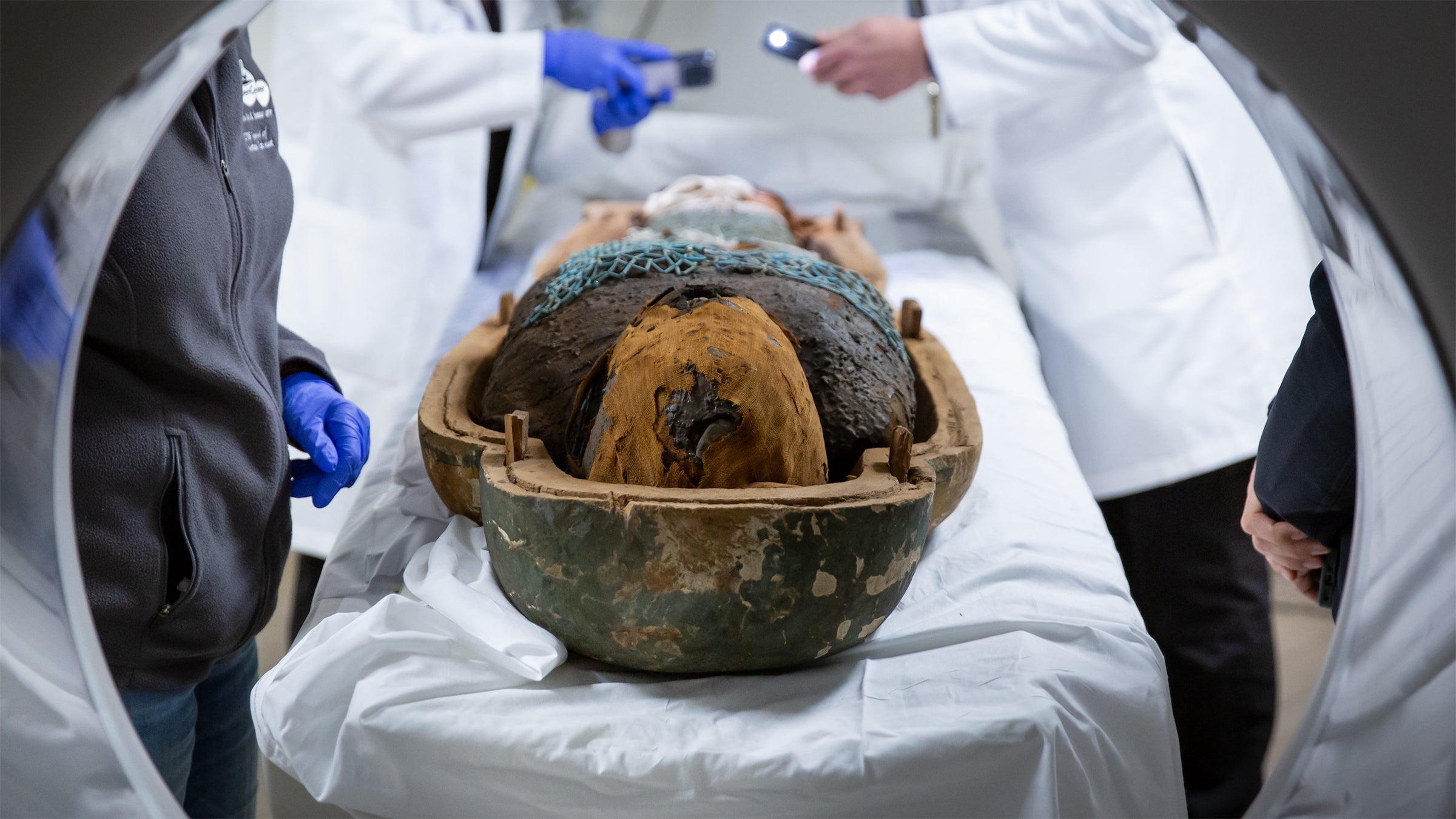
She didn’t have a lot of time: The mummies had been transported to the hospital from the California Science Center in Exposition Park, where they were to be displayed in Mummies of the World: The Exhibition when it opened Feb. 7. On the day of the CT scans, the mummies needed to be back at the California Science Center by 1 p.m. for installation into their displays.
That left Decker’s team just a few hours to glean knowledge concealed for more than 2,200 years. “I had my phone in my hand looking at the timing, making sure that we were capturing everything we could, because once they sealed them into that exhibit, they were off-limits to us,” Decker says. “This was our one shot at it.”
Ancient patients get a thorough exam
Decker is the 3D imaging lead with Keck Medicine of USC and founding director of the USC Center for Innovation in Medical Visualization, established in 2024 at Keck School of Medicine to push the boundaries of medical imaging and expand its role in patient care. The center uses advanced imaging technology and 3D visualization tools to create patient-specific 3D-printed models of body parts for complex surgical planning. Associate Director Jonathan Ford, associate professor of clinical radiology at Keck School of Medicine, is Decker’s longtime collaborator. The researchers hold joint appointments at Keck School of Medicine, the USC School of Cinematic Arts and the USC Viterbi School of Engineering, reflecting the interdisciplinary nature of their work.
Cutting-edge tools like high-resolution CT scans and 3D modeling aren’t just helping clinical radiologists to visualize patients’ illnesses and injuries with unprecedented precision. They’re also allowing USC researchers in various disciplines to reach across millennia in minutes, see the otherwise unseeable in rare artifacts without damaging them, and make profound discoveries about the past.
On that January morning, Decker inspected the mummies carefully before sending them one by one through the scanner. The top half of each mummy’s ornately painted wooden sarcophagus — whose symbols revealed each man’s name and details about his life — had been removed to facilitate the scans.
She was impressed by the handiwork that went into encasing the two ancient priests, part of a divine ritual that the ancient Egyptians believed would help ensure the deceased’s passage to eternal life. Nes-Min’s shroud was largely intact but showed some signs of wear around his face. A delicately beaded net covered the length of his shroud from chest to shins. Strands of colorful beads adorned the neck area.
Nes-Hor’s linen wrapping, blackened with age, was completely intact. Decker noticed a spot where the fabric had been tucked in to keep it in place.
“You think about somebody’s hands doing that,” she says. “It’s something that we all can relate to, of caring about someone, respecting someone enough to do that, to show that amount of love for them.”
For Decker, the scans were a way to extend that respect — to expose what lay beneath the mummies’ shrouds without unwrapping them or damaging them in any way.

In a typical CT scan of a living person, Decker takes around 1,000 images head to toe. “Each mummy got scanned at 25,000 images each,” she says. “We didn’t want to miss anything.”
Ford sat at a computer beside her transforming the images into 3D models of the mummies. When Ford visualized Nes-Min’s lumbar spine (lower back), the researchers saw a compression fracture in his L2 vertebra — an injury that would have given him intense back pain during his life.
Then they noticed something astonishing that prompted them to go back to the original images and closely reexamine them, slice by razor-thin slice.
A porthole to the past
Behind Nes-Min’s fractured vertebra was a small hole in the bone that could not have occurred naturally. When they zeroed in on the area, the researchers saw indications of tool marks and evidence of healing. The shape of the hole was consistent with trepanation, a surgical practice used in ancient Egypt in which holes were drilled into the skull to relieve pressure and pain.
The researchers realized they may have stumbled upon the first-known instance of ancient Egyptian spinal surgery. “We spent the next few weeks just staring at those images and sending them off to mummy specialists,” Decker says. “No one else has seen anything like that in the spine.”
Spotting the hole in Nes-Min’s vertebra opened a window into a rich set of human histories: the suffering of a priest with agonizing back pain; the caring hands of a practitioner who manipulated tools to attempt to provide relief; and a surgery that, if verified, will add to the annals of medical history.
Decker links these narratives to an objective she and Ford shared with Diane Perlov, senior vice president for special projects at the California Science Center, who commissioned the scans to be included as part of the exhibition. “Our goal was for modern people to remember that Nes-Min and Nes-Hor were living individuals walking this earth,” Decker says. “They had stories.”
“They’ve seen worlds we haven’t,” Ford says. “They’ve seen people and civilizations, had conversations, eaten food, laughed, joked.”
“This is part of our story as a species on this planet,” he adds.
A continuum of human innovation
Before Decker earned her doctorate in medicine, she studied forensic anthropology and trained under one of the world’s foremost mummy specialists. More than 15 years ago, when she was a graduate student at the University of South Florida in the same lab as Ford, Decker was involved in scanning mummies for the first iteration of Mummies of the World: The Exhibition when it debuted at the California Science Center in 2010.
Scanning Nes-Min and Nes-Hor, mummies never before seen in Los Angeles, was a full-circle moment for Decker, she says. The exhibition has traveled around the world since 2010; the current run in Los Angeles through Sept. 7 is its final iteration.

Perlov, an anthropologist, approached USC about scanning Nes-Min and Nes-Hor because the exhibition focuses on the science of mummies and mummification. “When visitors look at the CT scans, they’re scientists looking at data,” Perlov says. “They’re seeing something that only science can reveal.”
Imaging technology has advanced considerably since 2010 and even more so since the 1990s — the last time Nes-Min and Nes-Hor were scanned — allowing for more detailed and extensive findings.
Just as DNA analysis is now used to retrospectively solve crimes that occurred before the technology was available, Decker believes each new advance in imaging can offer a fresh perspective on ancient artifacts. “In many ways, we’re obligated to keep trying to figure out these mysteries and understand them using the tools that we now have,” she says.
Research by Lynn Dodd, professor of the practice of religion and spatial sciences at the USC Dornsife College of Letters, Arts and Sciences, centers on the integration of advanced visualization technologies into the study of the past. Director of the USC Archaeology Research Center and its Extended Reality Lab, Dodd employs a powerful micro-CT scanner at USC Viterbi to investigate how metal objects from the ancient Near East, Egypt and Mediterranean were made.
“The device lets us see minute spaces, including the microscopic bubbles that form when bronze is poured into a mold,” Dodd says. “It lets us get at action in the past that otherwise is invisible to us.”
Dodd has uncovered fabrication techniques that were innovative for their time. Much like Nes-Min’s apparent spinal surgery, these traces of human ingenuity remind us that our scientific and technological strivings are not so dissimilar from those of our forebears.
“We use innovation now to understand innovation then,” Dodd says.
Members of one human family
Decker recalls being amazed by the first images of Nes-Hor, who lived circa 190 BCE, that appeared on Ford’s screen. “You could see his ears, his eyelids, his eyes,” she says — a level of imaging detail not technologically achievable until very recently.
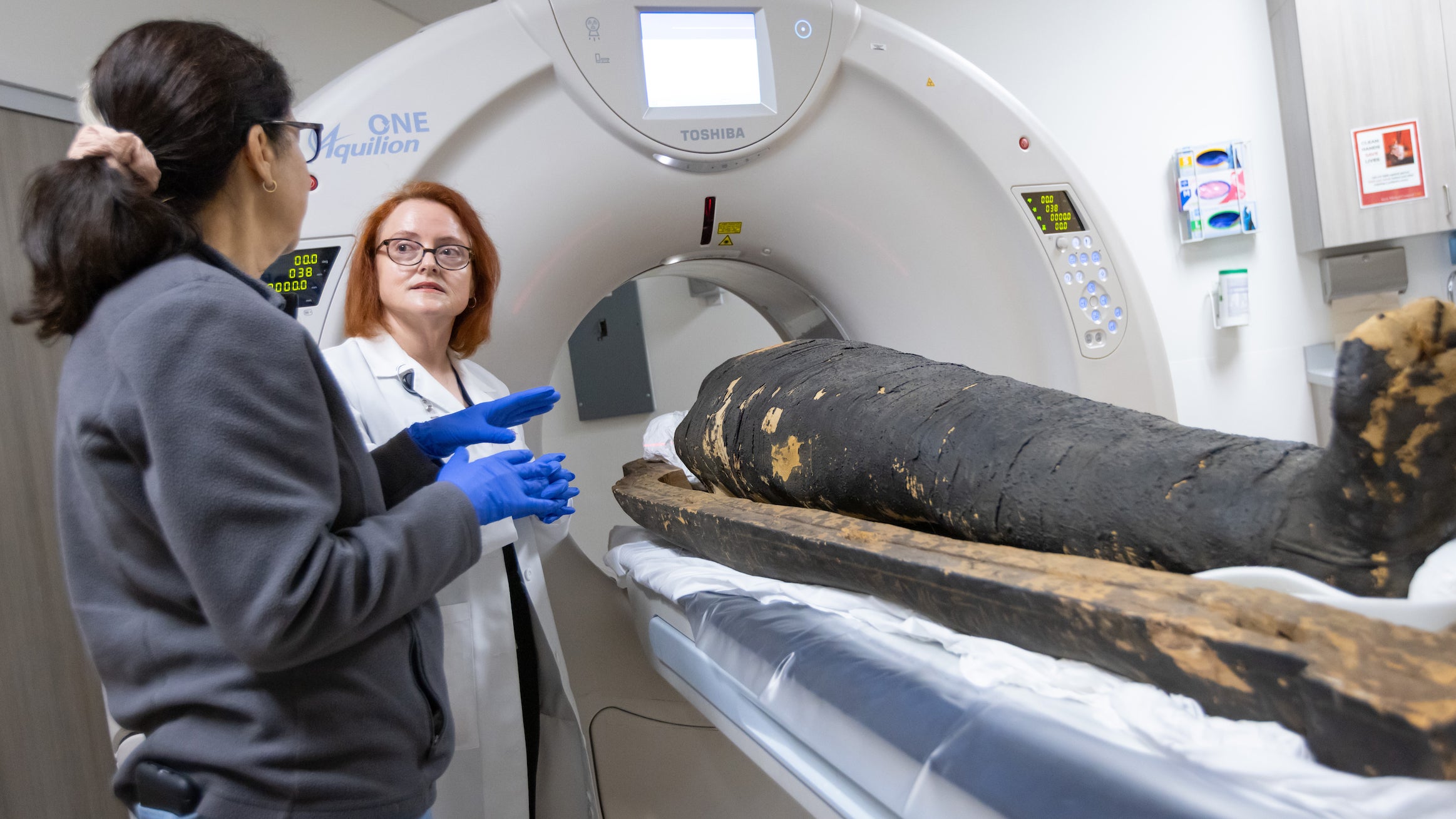
Perlov was in the control room with Decker and Ford during the scans. She marveled at how well-preserved Nes-Hor’s and Nes-Min’s facial features were. Though they lived over 2,200 years ago, their facial characteristics were indistinguishable from those of modern-day humans. “They looked so familiar,” Perlov says. “The detail and clarity were more than I expected. It was wonderful.”
As the CT scanner moved from the mummies’ heads to their toes, a host of discoveries emerged. Nes-Hor’s scans uncovered dental issues and a left hip fracture a few years before his death at age 60 that did not heal properly. The condition of the bone and muscle tissue surrounding the hip suggested Nes-Hor used his left leg despite the injury.
“This kind of fracture would have been very taxing on someone, especially back then, so he wouldn’t have been able to walk unassisted,” Decker says.
Previous scans of Nes-Min, who lived around 330 BCE, determined that he had probably died of a dental abscess around age 40. But the new high-resolution scans ruled out that possibility; his dental issues weren’t severe enough to be fatal. In addition to his spinal irregularities, other new findings included amulets placed on his head and chest representing scarab beetles and a fish. In ancient Egypt, such amulets were thought to protect the deceased on their journey to the afterlife.
“The mummies look like very exotic specimens, but once you see beneath the surface, and you see that they suffered from lower back pain and toothaches and had a broken bone or two, you see them as real people that had a lot of the same ailments that we did,” Perlov says. “It humanizes them.”
Holding history in your hands

Just as they do for Keck Medicine of USC patients and surgeons, Decker and Ford printed out life-size reproductions of the mummies’ injured body parts, including Nes-Min’s fractured spine and Nes-Hor’s damaged hip joint, using medical-grade 3D printers housed in the USC Center for Innovation in Medical Visualization. They also printed replicas of Nes-Min’s amulets.
When the prints of the amulets were complete, Decker turned them over in her hands. Though they were just facsimiles, their effect on her was akin to the real thing. “This is the first time in 2,300 years that someone is holding these,” she thought.
Now on display alongside the mummies and their scans at the California Science Center, the reproductions allow visitors to make a tactile connection with ancient history.
“If we are feeling something embodied — feeling it, looking at it, seeing it all around us — we have an emotional reaction,” Dodd says. “And we know that we learn best when things are connected with emotions.”
At the USC Archaeology Research Center’s Extended Reality Lab, Dodd oversees projects that use 3D VR tools to allow users to virtually touch historical artifacts that would otherwise be out of reach. These include a 15th-century illuminated manuscript and objects inside the oldest hunting lodge on Catalina Island.
“3D technology helps to connect people in a visceral, embodied, exciting way to places and stories about the past,” she says.
Inside Mummies of the World: The Exhibition, that sense of emotional intimacy with bygone eras is palpable. “Visitors are just in awe,” Perlov says. “The reaction is whispered wonderment. Science makes these discoveries possible, but it’s this emotional connection that makes science memorable.”
As detailed as Decker and Ford’s scans and reproductions of the mummies are, history has proven that innovation is a human constant. Decker knows that one day the work will be outdated by further breakthroughs in imaging — perhaps even some made by herself and her USC collaborators.
“The methods we used are the best available to us today,” she says. “There’s something new coming out next year or the year after that, and it’s going to change everything.”
_______________
USC faculty and staff can explore Mummies of the World: The Exhibition at the California Science Center and save on admission with promo code “MYSTERY.” Discounts are valid online on the California Science Center website through Sept. 7, 2026.


