Cell by cell: Rebuilding the body
USC researchers are revolutionizing how we treat disease by harnessing stem cells as “living medicine.”
Anyone who’s healed from a cut or a scrape has witnessed the incredible regenerative power of stem cells. These cells can create identical copies of themselves, creating new cells and tissues that replace damaged ones.
Stem cells are active in some areas of our body throughout our lives, like the skin and blood. But in many critical organs, including the heart and kidneys, stem cells are absent. When such tissues are damaged due to aging, injury or disease, they don’t regenerate, leading to devastating health consequences.
USC researchers are at the forefront of an emerging field called “clinical regenerative medicine,” which taps stem cells’ restorative powers to tackle some of the hardest-to-treat diseases, ranging from heart failure to blindness.
“We now have the ability through stem cells to generate replacement cells that we can use as therapeutics to rebuild the human body,” says Charles (Chuck) Murry, a renowned expert in regenerative heart medicine. In August, Murry joined Keck School of Medicine of USC as the new head of USC Stem Cell, chair of the department of stem cell biology and regenerative medicine, and director of The Eli and Edythe Broad Center for Regenerative Medicine and Stem Cell Research.
Launched in 2013, USC Stem Cell is a universitywide initiative that connects over 100 research and clinical faculty in multiple disciplines across USC and Children’s Hospital Los Angeles (CHLA) with the common goal of translating basic stem cell science into clinical therapies. It has matured over the past decade with support from the Eli and Edythe Broad Foundation as well as the California Institute for Regenerative Medicine (CIRM), a state organization created to accelerate stem cell research.
USC Stem Cell collaborators are employing stem cells to grow organ and tissue replacements, halt or reverse the progression of life-threatening diseases and create living models of human organs in the lab, providing novel platforms to screen for disease-fighting drugs.
“Clinical regenerative medicine is going to have an impact on par with antibiotics or vaccinations,” Murry says. “It’s going to be revolutionary.”
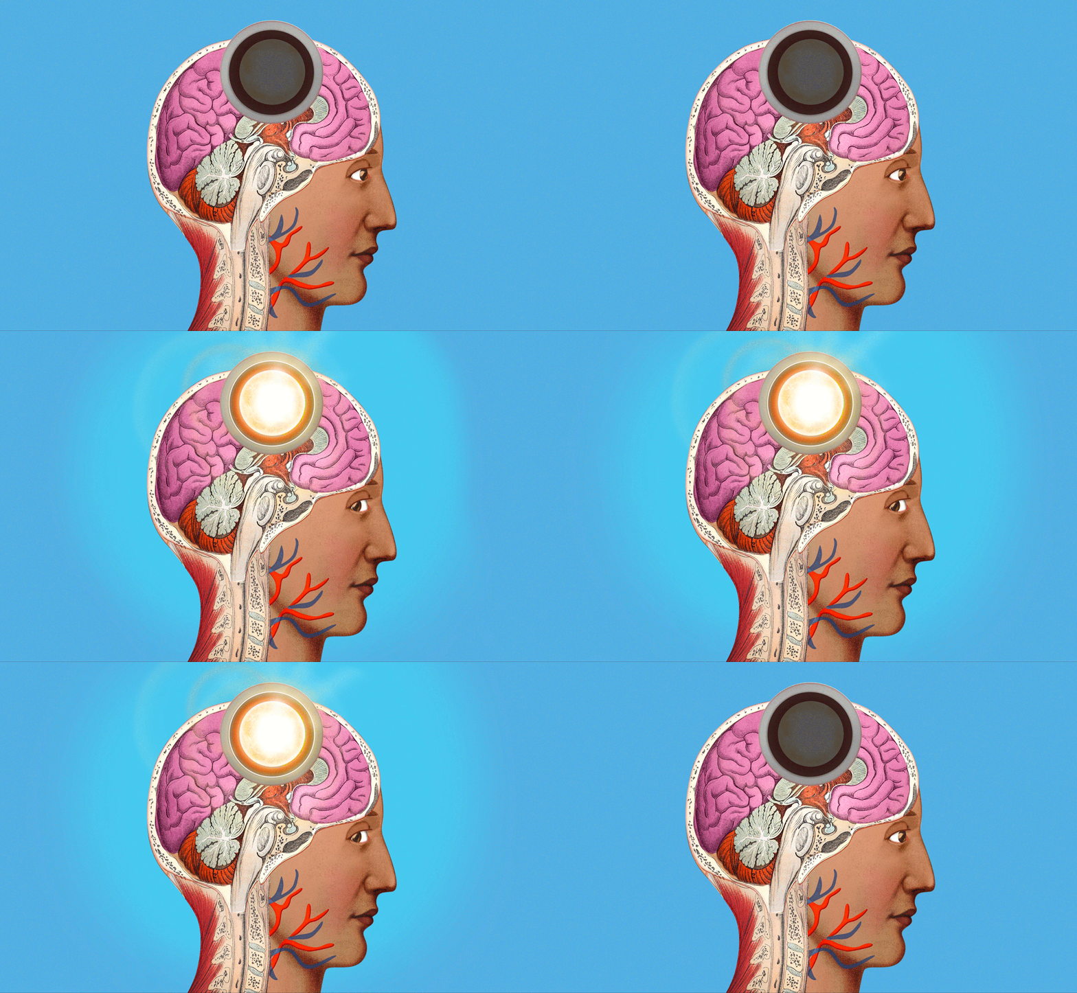
The stem cell projects currently underway across USC will transform treatments from our skull to our knee and many organs in between. As with the classic board game Operation, we’ve broken down a selection of these projects by body part — funny bone not included.
SKULL
The bones of our skull protect one of the most important human organs: the brain. When those bones are compromised either because of a congenital condition, injury or surgery, the defect may affect not only the appearance of the skull but also brain function itself. Since skull bones do not regenerate after infancy, surgery is often needed to repair defects.
Yang Chai, University Professor, interim dean and associate dean of research at the Herman Ostrow School of Dentistry of USC, treats craniofacial birth defects. His work inspired him to develop an innovative treatment option for patients with significant defects — essentially holes — in the calvarial bones at the top of the skull.
Typically, these holes are surgically closed with a metal or plastic plate. For pediatric patients, neither option is ideal because a synthetic plate will not grow in concert with the child’s developing brain. Many of these implants fail within 20 years.
Collaborating with Yong Chen, professor of aerospace and mechanical engineering and industrial and systems engineering at the USC Viterbi School of Engineering, Chai designed a “living” implant for skull bone regeneration. It’s a 3D-printed scaffold made of a mineral found in teeth and bones called hydroxyapatite. The scaffold can be customized to fit a patient’s defect precisely and seeded with a patient’s own bone marrow-derived stem cells.
In animal models, as the stem cells mature, they grow and integrate with the surrounding skull to cover the defect within about six months. The new bone becomes as strong as the native bone. Chai and his collaborators — including Mark Urata, professor of clinical surgery at the Keck School of Medicine of USC — are poised to begin clinical trials in humans.
“Ours is going to be a biological solution for a biological problem, instead of a mechanical solution [i.e., a synthetic plate] for a biological problem,” Chai says.
BRAIN

Studying human brain development is important in understanding how neurodevelopmental issues such as autism spectrum disorder arise and progress. But traditional research methods have proved insufficient for observing the human brain as it develops. Current brain imaging techniques don’t offer a resolution that is high enough to understand brain functionality at the cellular level. Postmortem human brains don’t provide a window into early growth, and animal models don’t possess the complexity of the human brain.
“The best way to access human brain development is really to take advantage of human stem cells,” says Giorgia Quadrato, assistant professor of stem cell biology and regenerative medicine at the Keck School of Medicine of USC.
Quadrato has pioneered a way to grow masses of brain cells in the lab from stem cells. Within six months, these cells mature into tiny structures (“brain organoids”) about two millimeters in diameter that contain all the various cell types of specific brain regions. The organoids allow Quadrato’s team to study the cells’ development in real time, model neurodevelopmental diseases and screen for potential new treatments.
One of her lab’s most noteworthy innovations is an organoid model of the cerebellum, a brain region that plays a role in movement, cognition and emotion. In certain neurodevelopmental and neurodegenerative disorders like autism spectrum disorder and cerebellar ataxia, cells in the cerebellum called Purkinje neurons degenerate. Quadrato’s team is the first to succeed in growing Purkinje cells with all the features of functional neurons in a human system.
While the cerebellum models were grown from healthy cells, Quadrato has also grown organoid models of the cortex — a brain region involved in cognitive function — derived from the cells of a patient with a disease-causing variant of the SYNGAP1 gene. This variant is one of the top risk factors for autism spectrum disorder. The lab’s organoid models shed new light on how the variant disrupts early development of the cortex.
EYES
As we age, changes to the retina, the delicate tissue at the back of our eyes, can lead to vision loss. Photoreceptor cells that sense light and send signals to the brain can deteriorate due to inflammation, genetics and lifestyle habits such as smoking. At the center of the retina, the macula, which controls straight-ahead vision, is particularly vulnerable to damage.
Age-related macular degeneration (AMD) is the leading cause of severe vision loss among people over 50 in Western countries. Because there’s no known way to regenerate photoreceptors, AMD has had no effective treatments — until now.
Mark Humayun, University Professor of ophthalmology at the Keck School of Medicine of USC and co-director of the USC Roski Eye Institute, and his collaborators are using stem cells as a gateway to restoring vision in people with dry AMD, the most common form.
The disease affects not only photoreceptor cells but also a vital group of cells that protect them from damage called retinal pigment epithelium (RPE) cells. Humayun and his team developed a tiny patch filled with RPE cells derived from stem cells that can be surgically implanted in the eye, where the young cells can shield patients’ photoreceptors from further degeneration.
Clinical trials of the patch are run by Regenerative Patch Technologies, the company he co-founded with Dennis Clegg and the late David Hinton. In the first phase of the trial, 27% of patients experienced improved vision.“We found that these young RPE cells are very robust,” Humayun says. “They survive much better than adult-aged RPE. Patients who improved are still keeping the same level of vision three years later.”
This fall, during the trial’s second phase, the patch will be tested in a larger number of dry AMD patients. Among them will be patients with less severe disease than patients in the first phase. Humayun expects that in this phase the patch will improve vision in even more patients and to a greater extent.
MOTOR NEURONS
Amyotrophic lateral sclerosis (ALS) — often called Lou Gehrig’s disease — is a devastating neurodegenerative disease with no known cure. It affects motor neurons, which are nerve cells in the brain and spinal cord that control voluntary muscle movement and breathing. Over time, as motor neurons degenerate, the brain can no longer command muscle movements, and people with ALS may lose their ability to speak, eat, walk and breathe.
Finding universally effective treatments has been difficult because the disease’s root causes are not fully understood. While having a family member with ALS increases your risk of developing it, most cases occur with no family history or clearly associated risk factors. “In these cases, we believe that multiple gene mutations come together in one person,” says Justin Ichida, an associate professor of stem cell biology and regenerative medicine at the Keck School of Medicine of USC whose research is centered on fighting the disease.
Stem-cell technology has provided Ichida an avenue for new leaps in discovery. He and his team reprogram adult skin or blood cells from an ALS patient into stem cells. These cells are then coaxed into developing into motor neurons that are genetically identical to the patient’s own motor neurons. The process has allowed Ichida’s lab to test thousands of compounds to see how the neurons respond.
One of Ichida’s most exciting discoveries is that inhibiting a protein called the PIKFYVE kinase improves the survival of motor neurons derived from patients with varying types of ALS. His startup company, AcuraStem, developed a druglike molecule to suppress this kinase and licensed it to a pharmaceutical company, which is now developing it into a drug for clinical trials. “This could be applicable for essentially all ALS patients, rather than just a subset that have a certain type of mutation,” Ichida says.
EARS

Inside your cochlea — the spiral structure inside your inner ear — a few thousand tiny sensory receptors with bundles of hairlike protrusions (so-called “hair cells”) perform a function essential for hearing. They convert the mechanical vibration evoked by sound waves into electrical signals that your brain interprets. When they’re damaged either from wear and tear or disease, hair cells can’t be normally replenished.
That’s true in humans, mice and other mammals. But in non-mammalian vertebrates, such as songbirds, damaged hair cells regenerate in a matter of weeks. The difference comes down to the activity of stem cells called supporting cells.
“The first thing a supporting cell needs to do is sense that there is an injury and divide into progeny that will mature into hair cells,” says Ksenia Gnedeva, assistant professor of otolaryngology – head and neck surgery and stem cell biology and regenerative medicine at the Keck School of Medicine of USC. “That doesn’t happen in mammals. The very first step is blocked.”
Gnedeva discovered that a molecular cascade called the Hippo signaling pathway acts like a brake on that first step in regeneration in mammals. Then, she identified a small molecule that blocks the main enzyme in the pathway called the LATS kinase that can switch the cascade from a brake to an accelerator.
“When we applied the kinase inhibitor to inner ear sensory organs from mice, we saw something that had never been published before,” Gnedeva says. “All of the supporting cells were now able to reenter the cell cycle,” meaning that they began actively dividing in the petri dish. Remarkably, some of them grew into hair cells.
Gnedeva’s lab is now investigating how to effectively deliver this compound into the inner ear to restore hair cells in living mice. The hope is that the therapy may one day work to regenerate hair cells in humans and restore hearing to those with severe hearing loss.
KNEES
In your joints, a strong, flexible connective tissue called cartilage provides padding between bones and acts as a shock absorber and lubricant. Osteoarthritis occurs when the cartilage is compromised due to injury or aging, causing the bones to rub against each other, often painfully.
Very few stem cells are present in adult cartilage, meaning the tissue doesn’t naturally regenerate. Historically, treatments for osteoarthritis have been limited to painkillers and invasive surgery to replace damaged joints. But Denis Evseenko — professor of orthopedic surgery, stem cell biology and regenerative medicine, vice chair for research of orthopedic surgery and director of skeletal regeneration at the Keck School of Medicine of USC — and his collaborators have innovated new stem-cell-based treatments that promise to make osteoarthritis pain a thing of the past.
One is a “regenerative pouch” to replace cartilage that’s been damaged by a fall, sports injury or other trauma. The pouch, which is being developed by Evseenko’s startup company Plurocart, contains hundreds of thousands of young cartilage cells derived from stem cells. “It’s a little reparative structure that you can surgically deliver right into the cartilage defect,” Evseenko says.
In animal models, the pouches have shown phenomenal results: The juvenile cells grow into mature cartilage, preventing osteoarthritis. Evseenko is now planning the first clinical trial in humans with knee injuries.
Evseenko’s lab has also created a therapy for age-related osteoarthritis that is being developed by his startup company CarthroniX. As we age and cartilage is progressively lost, painful inflammation can set in. “Chronic inflammation is a signal for local stem cells that it’s a bad environment and they should not build new tissue,” Evseenko says.
He identified a compound that disrupts a key immune receptor’s over-activation due to inflammation. When this compound is injected into the knees of patients with mild to moderate osteoarthritis, inflammation is suppressed, stem cells are activated to slowly regrow tissue and pain is significantly reduced.
HEART

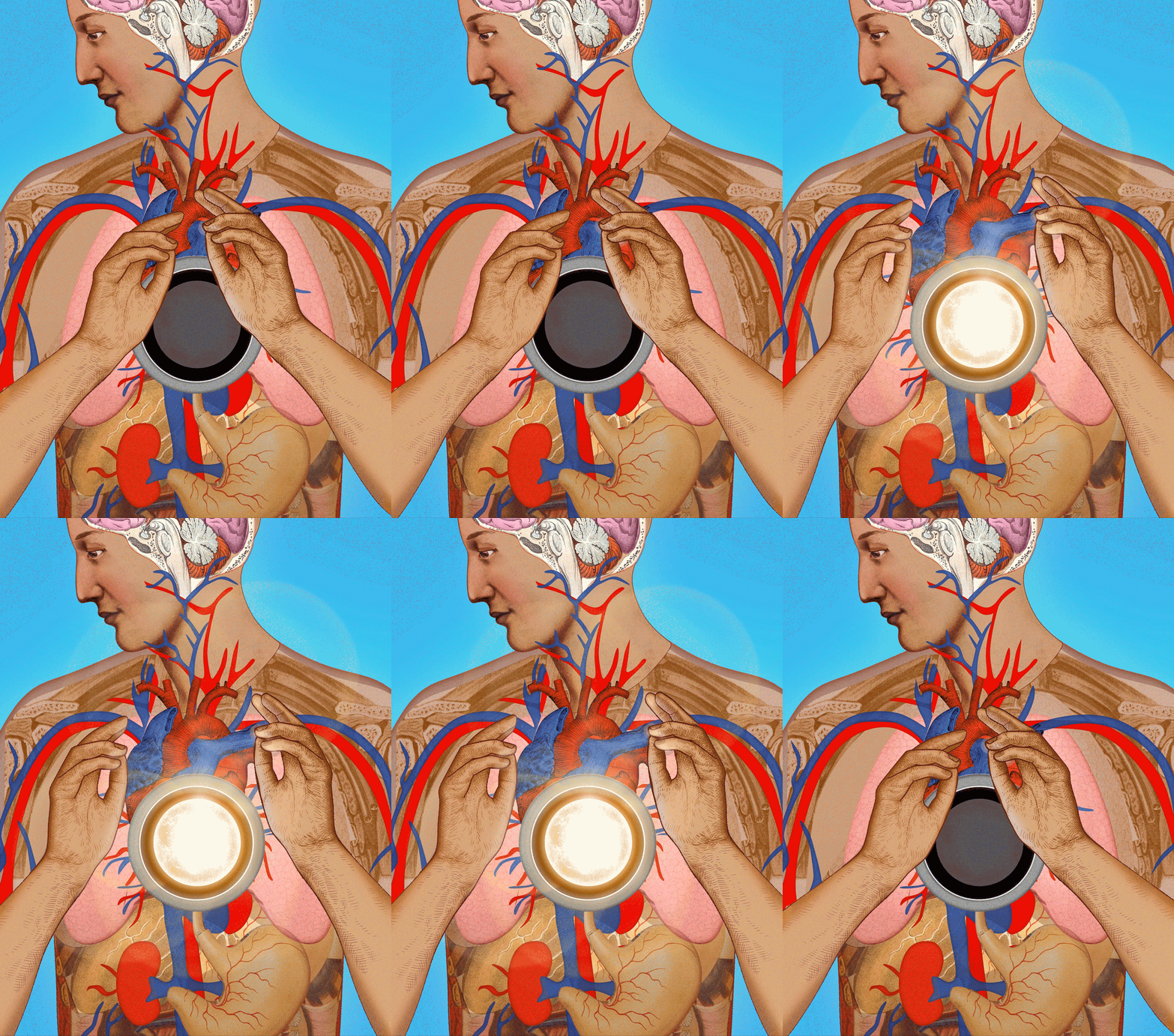
After a heart attack, many people go on to lead productive lives — but the heart muscle itself is forever changed. Since adult heart cells do not regenerate, the scar tissue formed in areas of the heart attack can compromise the heart’s ability to pump. Over time, reduced function can lead to heart failure, depending on the amount of scar tissue and the size of the heart attack.
Murry, the head of USC Stem Cell, has discovered a way to use heart cells derived from stem cells to strengthen hearts damaged by scar tissue. “By transplanting these heart muscle cells, we can rebuild the heart and restore its contractile function,” Murry says.
He likens the process to growing a garden: The injured heart becomes the soil where stem-cell-derived heart cells are planted. The “roots” are the connective tissue and blood vessels that the new cells call in to feed them. Murry anticipates that clinical trials of the transplanted cells will begin within the next couple of years.
Megan McCain, associate professor of biomedical engineering at the USC Viterbi School of Engineering and of stem cell biology and regenerative medicine at the Keck School of Medicine, has innovated complex living tissue models from human stem cells that allow her to visualize heart scar tissue formation in real time.
Her team seeds quarter-size silicone wafers with stem cell-derived heart muscle cells. As the cells grow, they align and begin to beat together much as they would in an actual human heart. McCain can then induce what she calls a “heart attack on a chip” — by selectively depriving some of the cells of oxygen — and observe how the cells remodel themselves after the injury.
“The next step is to develop interventions to change how the heart heals itself,” McCain says, to reduce how much scar tissue forms in the first place.
Vaughn Starnes, chair and distinguished professor of surgery at the Keck School of Medicine and founding executive director of Keck Medicine’s USC Cardiac and Vascular Institute, is exploring stem cells as a treatment for children born with a congenital condition called hypoplastic left heart syndrome. In those affected, the left side of the heart is underdeveloped, leaving the baby with one, instead of two, functioning ventricles to pump blood. Over time, the single ventricle fails in 10% to 15% of patients.
Early evidence suggests that when stem cells from a baby’s own umbilical cord blood are differentiated into heart cells and injected into that ventricle, they strengthen the muscle and improve its function — potentially reducing the chances of failure down the line.
KIDNEY

Our kidneys are vital organs to sustain life. They filter waste products from our blood and produce urine we excrete. Because of the increasing prevalence of diseases that injure the kidneys, including hypertension and diabetes, chronic kidney disease is on the rise in the United States, affecting one in seven adults.
Kidney transplants can save lives — but the demand for donor kidneys far exceeds the supply. Each day, an estimated 13 Americans die waiting for a kidney transplant.
Zhongwei Li, assistant professor of medicine and stem cell biology and regenerative medicine at the Keck School of Medicine of USC, has pioneered a “synthetic kidney” using human stem cells that may soon provide an alternative to kidney transplants from a donor.
The architecture of a human kidney is complex. A million functional units called nephrons organize themselves into a treelike structure to drain the urine they produce. Once injured, nephrons can’t regenerate. Li developed a method for turning stem cells into kidney progenitor cells, the cell type necessary for kidney development in a human embryo.
“By using those building-block progenitor cells, we have successfully assembled a treelike structure with nephrons attached to the structure in a petri dish,” Li says.
His team has demonstrated that these “kidney organoids,” when transplanted into animal models, can grow into synthetic kidneys that produce urine just like natural kidneys. Li anticipates that within five to 10 years, the technology will be advanced enough for clinical trials in humans.
As he works with collaborators in fields including kidney development, 3D bioprinting and kidney physiology to perfect the synthetic kidney’s design, Li stresses that the power of his innovation comes from nature itself.
“Progenitor cells are hardwired to build a treelike structure,” Li says. “We direct the cells to do what they’re programmed to do.”
PANCREAS
For the more than 38 million Americans with diabetes, the disease significantly affects daily life and poses long-term health risks. While it can be managed with medication, diet and lifestyle, there’s currently no cure.
Diabetes occurs when your blood sugar is too high, either because your pancreas makes little or no insulin (Type 1 diabetes) or your cells don’t use insulin properly (Type 2 diabetes). In the latter condition, your pancreas may be producing insulin — just not enough to keep your blood sugar in the normal range.
“Whether it’s Type 1 or Type 2 diabetes, fundamentally you need more functional insulin cells,” says Senta Georgia, associate professor of pediatrics and stem cell biology and regenerative medicine at the Keck School of Medicine of USC. “For a number of years, my lab has been working on ways to make insulin cells from stem cells, trying to understand the barriers that exist and improve the processes that underlie how we make these cells.”
Georgia aims to support the development of clinical regenerative therapies for diabetes. These may one day include transplanting stem cell-derived insulin cells into the pancreas, coaxing a patient’s pancreas to make more of its own insulin cells or improving the function of existing insulin cells.
One of her lab’s current areas of focus is investigating why COVID-19 infections sharply increase the risk of developing Type 2 diabetes. Her team is using stem cell models to understand the virus’ impact on insulin cell function and survival, with an eye toward identifying and blocking pathways to injury.
They’re also collaborating with clinicians at CHLA to study the cells of pediatric patients with genetic forms of diabetes. A recent project illuminated the critical importance of a gene called NEUROGENIN3 in enabling stem cells to mature into insulin cells. Children with a mutation in this gene develop a severe form of diabetes.
CELLS
While many USC researchers are employing stem cells to regenerate damaged tissues and organs, Ian Ehrenreich — divisional dean for life sciences and professor of biological sciences at the USC Dornsife College of Letters, Arts and Sciences — is pioneering new ways to modify stem cells themselves.
Genetically modified stem cells hold great promise for therapeutic purposes. Ehrenreich points to one recently developed example: the first FDA-approved gene therapy for patients with sickle cell anemia, a blood disorder caused by a genetic mutation. In this therapy, the blood stem cells of patients are modified by genome editing and transplanted back into patients’ bone marrow, where they produce a protein that prevents the “sickling” of red blood cells.
“The type of genetic engineering involved in this therapy is simple,” Ehrenreich says. “They’re breaking one genetic element. But a lot of things we’re going to need to do [to develop new stem-cell therapeutics] are going to require more sophisticated genetic engineering. That’s the realm where my lab works.”
He and his team have developed new techniques for building large pieces of DNA (synthetic chromosomes), involving less time and cost than ever before possible. “These big DNA constructs will be important for reprogramming stem cells to serve particular tasks,” Ehrenreich says. DNA synthesized using these techniques could also be added to a stem cell to compensate for genes that a patient is missing.
One of Ehrenreich’s intracampus collaborations is with Leonardo Morsut, assistant professor of stem cell biology and regenerative medicine at the Keck School of Medicine of USC. Morsut engineers stem cells to divide in particular ways to grow tissues of desired structure and function. Employing large synthetic chromosomes from Ehrenreich’s lab “might enable more complex programming of these cells, which could be beneficial in eventually making [synthetic] organs,” Ehrenreich says.


