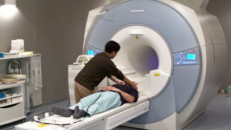
Functional MRI scanners provide maps of signals in the brain, but scientists know these maps may be slightly inaccurate. (Photo/Janne Moren)
Rare eye condition gives scientists a chance to improve brain scan studies
A student’s unusual vision issue gives USC researchers an opportunity to calculate a relationship between neural activity and the blood
There is an elephant in the room each time scientists conduct a brain scan study.
Functional MRI scans provide colorful maps of signals in the brain, but scientists know these maps may be slightly inaccurate. That’s because the scanner isn’t tracking neural activity directly. The scanner is measuring changes in the brain’s blood oxygenation level.
Scientists have been running bran scan studies with the assumption that the stimuli they use in their experiments prompts neurons to respond and in turn, changes the blood flow in the vessels that supply these neurons. However, scientists don’t know with any mathematical precision how the blood’s response relates to neuron activity.
“We’ve used this method for the last 25 years, and we don’t actually know the exact relationship between the blood signal that we see and the neural signal that we want to infer,” said USC neuroscientist Bosco Tjan.
The missing mathematical link
Tjan, a professor of psychology at the USC Dornsife College of Letters, Arts and Sciences and co-director of the Dornsife Cognitive Neuroimaging Center, said scientists have attempted to quantify the relationship between the blood oxygenation — known as a “hemodynamic” response — and neural signals.
For example, scientists at one point surgically inserted an electrode in a monkey’s brain, he said. The device gave them a direct measure of neural activity while the monkey was being scanned. But the study had significant shortcomings: It was invasive, and it could not be tried on human subjects.
Tjan said an opportunity was presented to him that could lead scientists to the missing mathematical link between the neural activity and blood response — without having to conduct any sort of invasive surgery.
A student interested in working in his lab had a rare vision problem — “achiasma” — a condition that allowed Tjan and his team to test and discover the mathematical relationship between neural and blood responses.
People who have achiasma, a congenital condition, typically have modest vision loss. However, the nerve connections between their eyes and the brain are far from normal. The condition is rare and likely under-diagnosed because it can only be detected with a brain scan — a tool which most optometrists who conduct routine eye checkups do no have.
Ordinarily, the eyes work like cameras, recording the scene before them. Each eye is recording the scene in two halves — the left and the right visual fields. Inside, neurons transmit the visual information along the optic nerves into the brain. The scene in the left visual fields of both left and right eyes is carried to the right brain hemisphere for processing. At the same time, the scene in the right visual field of both eyes is carried to the left brain hemisphere.
3-D viewing
When the left and right eyes’ images of the same scene are combined in the brain, the person can see in three dimensions.
The world looks flat to achiasma patients because they have a faulty wiring in the optic nerves. Instead of bringing images of the same visual field from both eyes to the same brain hemisphere so that the images can be combined, the miswired nerve fibers carry images from both visual fields of the same eye to the same brain hemisphere. This means the images from the two eyes end up in separate hemispheres and are never combined.
Neurons in the brain that are receiving input from the left visual field do not interfere with neurons receiving input from the right visual field because if they did, scenes from left and right visual fields would obscure each other.
However, because of the faulty wiring in achiasma, these neurons are located very closely to one another in the brain, so much so that they share the control of the local blood supply. This very unusual arrangement of noninteracting neurons enabled Tjan and his team to test, through a series of brain scans, whether the relationship between the neural and blood responses is linear, as scientists had long assumed.
[The research] opens up a new avenue for scientists to address the basic question of what functional MRI measurements really mean
Bosco Tjan
They found it is not. The functional MRI signal magnitude is proportional to roughly the square root of the underlying level of neural activity.
“We found the missing mathematical link between neural and blood responses from a unique individual, and it did a good job in accounting for other data in the literature,” Tjan said. “It opens up a new avenue for scientists to address the basic question of what functional MRI measurements really mean.”
His team is now developing new analytical techniques to provide a better interpretation of functional MRI signals.
Study co-author and USC psychology researcher Christopher Purington said that while more study is needed, “the exciting thing about this study is it brings us closer to what we are actually trying to study with fMRI — the neural activity.”
The team’s results were published recently in the journal eLife. Pinglei Bao, a former USC graduate in Tjan’s lab, is the lead author.


