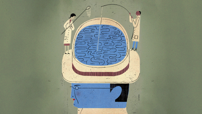
(Illustration/David Plunkett)
Brilliant minds, healthy brains
USC brain researchers are finding novel ways to image, detect and treat diseases.
This spring, scientists from the Keck School of Medicine of USC opened a new window into understanding the brain — literally.
Thirty-nine-year-old Jared Hager had injured his brain in a skateboarding accident. During emergency surgery, half of Hager’s skull was removed to relieve pressure on his brain, leaving part of the organ covered only with skin and connective tissue. A team at the Keck Medical Center of USC reconstructed his skull using a custom prosthesis that contained a transparent window.
The window, designed in collaboration with colleagues at California Institute of Technology, allowed the researchers to evaluate Hager’s brain function in a remarkable new way. While Hager played video games and strummed a guitar, the research team collected high-resolution brain data using functional ultrasound imaging. This type of imaging reveals brain changes that occur when a patient is performing a task — information that can be critical for assessing and treating traumatic brain injury.
“Functional ultrasound imaging can’t be done through the skull or a traditional prosthesis,” says Charles Liu, a professor of clinical neurological surgery, urology and surgery at the Keck School of Medicine and director of the USC Neurorestoration Center, who led the research team. “This is the first time physicians have been able to do it noninvasively in an awake patient through a window. The window allows us to monitor brain function and guide treatment in ways that were not possible before,” he says.
Effective treatment options for brain injuries and diseases have long been elusive. That’s in part because the brain — the command center of thinking, sensing, movement and emotion — is so complex to understand. The brain is also challenging to observe and study, especially in living humans.
Liu’s team at the USC Neurorestoration Center, which specializes in developing novel strategies to restore neurological function in those with injured or diseased nervous systems, is one of the many research groups at USC working to address the formidable challenges presented by brain diseases, from traumatic brain injuries and epilepsy to Alzheimer’s disease and other forms of dementia. Using advanced technologies and methodologies, they’re finding revolutionary ways to bring new clarity to the mysteries of our gray matter.
Alzheimer’s disease is one of the most enigmatic brain afflictions and among the greatest health care challenges facing the nation. It affects nearly 7 million Americans — a number expected to double by 2060 — and there’s no known cure.
MAPPING THE BRAIN
Alzheimer’s disease is one of the most enigmatic brain afflictions and among the greatest health care challenges facing the nation. It affects nearly 7 million Americans — a number expected to double by 2060 — and there’s no known cure.
The disease is characterized by two hallmark changes in the brain: plaques made of a protein called beta-amyloid and tangles made of a protein called tau. Scientists have yet to discover what causes these proteins to accumulate. Some have speculated that dysfunction in the blood-brain barrier (a membrane that keeps harmful substances in the blood from reaching the brain) and inflammation of the brain’s blood vessels may set the stage for protein buildup.
Arthur Toga — Provost Professor of ophthalmology, neurology, psychiatry and the behavioral sciences, radiology and engineering and the Ghada Irani Chair in Neuroscience at the Keck School of Medicine — has developed cutting-edge imaging techniques that offer new insight into these parts of the brain.
At the Laboratory of Neuro Imaging (LONI) in the USC Mark and Mary Stevens Neuroimaging and Informatics Institute, Toga and his research team program the radio frequency pulses used in magnetic resonance imaging (MRI) to observe the blood-brain barrier and the fluid-filled spaces around the brain’s blood vessels.
“These innovative techniques are really improving our ability to look at the most minute features of brain organization and brain function that may be affected by this disease process,” Toga says.
As Toga’s team works to create the most detailed neurological maps in existence, they’re also adding to LONI’s Image and Data Archive, a tool developed by Toga and his collaborators to facilitate real-time data sharing among thousands of researchers worldwide. Toga believes that such cross-institutional collaboration is essential for solving the riddle of Alzheimer’s.
He is one of the principal investigators on the Health and Aging Brain Study – Health Disparities (HABS-HD), a joint effort among five institutions to address the lack of diversity in Alzheimer’s disease research. Hispanic and African American populations experience a significantly greater risk of developing Alzheimer’s disease than non-Hispanic whites, yet much of what is known about the disease is based on data gathered among the latter group. HABS-HD has enrolled thousands of participants from underrepresented groups and is generating the world’s largest repository of data describing these populations.
“The path to discovery is paved with data,” Toga says.
PREVENTION IN PILL FORM
Thanks in part to the Image and Data Archive, the first drug to slow progression of Alzheimer’s disease — lecanemab — came on the market last year.
Pharmaceutical companies used data from the archive to develop the drug and design clinical trials. Those trials have shown that lecanemab, which targets and removes abnormal beta-amyloid deposits in the brain, slows down declines in memory and thinking by about 30% in those with early-stage Alzheimer’s.
For Paul Aisen, professor of neurology at the Keck School of Medicine and the founding director of the Alzheimer’s Therapeutic Research Institute, 30% improvement is not enough. “We’re very focused on research to find the best drugs to do better,” says Aisen, who leads research into evaluating drugs to treat — and even prevent — the disease’s underlying pathology in the brain.
Lecanemab is approved for people who have a confirmed diagnosis of Alzheimer’s disease in its mildest symptomatic stages. Aisen’s team is investigating the drug’s potential use in those whose brains are beginning to show Alzheimer’s changes but do not yet have any symptoms. “We believe if we remove the beta-amyloid as it’s starting to accumulate, while the brain is still functioning normally, we’re going to have an impact that is much better than a 30% slowing of disease progression,” Aisen says.
Another of the institute’s projects focuses on using lecanemab in conjunction with drugs that target brain tangles made of tau. By targeting both tau and beta-amyloid deposits at once, Aisen’s team aims to put the brakes on the disease even more effectively.
Aisen believes that within a decade, such pharmaceutical advances may make Alzheimer’s disease a thing of the past. He sees a future where clinicians will monitor everyone starting in middle age, identifying those who are at risk for the disease and prescribing drugs that can keep proteins from accumulating abnormally in the brain.
“It will be like checking your cholesterol and treating high levels in midlife so that you don’t get heart attacks and strokes in later life,” he says. “We think we can prevent Alzheimer’s disease the way statins [drugs that lower cholesterol] have dramatically lowered the occurrence of heart attacks.”
“We think we can prevent Alzheimer’s disease the way statins have dramatically lowered the occurrence of heart attacks.”
Paul Aisen
BESPOKE BRAIN FITNESS
While Aisen envisions routine Alzheimer’s prevention for all, researchers at the USC Center for Personalized Brain Health at the Keck School of Medicine are focusing their prevention efforts on a subset of people who are known to have a high risk of developing the disease: those who are carriers of a fairly common genetic variant called APOE ε4.
Roughly one in four people carry one copy of the gene, elevating their risk of Alzheimer’s. Those who have two copies of the gene — 2% to 3% of the population — face eight to 12 times the risk for the disease.
“When people take a genetic test and find out they have the APOE ε4 gene, they are often scared and unsure of what to do — especially if they have a family member who already has dementia,” says Hussein Yassine, professor of neurology and gerontology at the Keck School of Medicine and director of the center. “We’re trying to fill this gap by providing resources to help patients do what they can to prevent the disease.”
The center has two components that inform one another: a clinic that develops personalized diet and exercise interventions for each patient to potentially slow cognitive decline, and a research wing focused on the development of new drugs. “This bridge between research and the clinic is quite novel,” Yassine says.
One example of the center’s translational approach is a multi-pronged investigation into the role of omega-3 fatty acids, which are found in foods like salmon and walnuts, in the brain health of APOE ε4 carriers. Imaging studies have shown that the brains of APOE ε4 carriers are deficient in omega-3s years before Alzheimer’s brain changes set in. Yassine launched a clinical trial to test whether early omega-3 supplementation in people with APOE ε4 can slow down disease progression. He also partnered with Kai Chen, professor of research radiology at the Keck School of Medicine, to invent a new imaging technique that traces omega-3s in the brain.
AI SEE YOU
High-resolution imaging techniques like those used by Liu, Toga and Yassine help researchers visualize the intricacies of brain tissue in never-before-seen ways. Yet the human eye itself has limitations that affect how brain images are interpreted. Andrei Irimia, associate professor of gerontology at the USC Leonard Davis School of Gerontology, is using artificial intelligence to push past those limits.
Irimia and his colleagues use an AI technology called deep neural networks to analyze MRI brain scans. The AI model allows Irimia’s team to identify subtle patterns in the brain scans that the human eye might not be able to detect.
One application of the technology is assessing biological brain age, an important factor in the development of Alzheimer’s disease and other forms of dementia. While the risk of developing these neurodegenerative diseases increases with age, not everyone’s brain ages at the same rate. “A person who is very fit and has a healthy lifestyle might experience low and relatively much slower rates of atrophy in the brain compared to more sedentary individuals,” Irimia explains.
He notes that traditional measures of brain aging, which include judging brain age by the thinning of the cerebral cortex, may not offer the fullest picture.
“By identifying patterns in a very large array of changes pertaining to brain anatomy, these deep neural networks can estimate brain age a lot better than we could based on measures that have been identified by humans,” Irimia says.
“A person who is very fit and has a healthy lifestyle might experience low and relatively much slower rates of atrophy in the brain compared to more sedentary individuals.”
Andrei Irimia
THE BRAIN ELECTRIC
At the Keck Medical Center of USC, where Liu and his colleagues implanted the transparent skull prosthetic, digital technologies are being integrated into the brain itself to help restore function in those with brain and spinal cord injuries.
In collaboration with colleagues at Caltech and the University of California, Irvine, Liu’s team is developing brain-computer interfaces to help paraplegic patients regain feeling in their legs and walk again. These interfaces offer a bidirectional communication link between the brain’s electrical signals and a bionic bodysuit (aka “robot exoskeleton”) worn by patients that can aid in movement.
Patients control the movement of the exoskeleton with their thoughts. Electrodes implanted in the brain record electrical impulses that orchestrate movement, which are then translated into commands that control the robotic suit. Not only do patients move, they can feel the motion. “When the robot exoskeleton moves, patients feel every step because key areas of the brain are stimulated,” Liu says.
Implantable brain devices are also being used for epilepsy treatment at the center. Responsive neurostimulation (RNS) implants monitor waves in parts of the brain where seizures begin, detect unusual electrical activity that can lead to a seizure and, within milliseconds, deliver small bursts of electrical stimulation to “stop the seizure in its tracks,” says Christianne Heck, professor of clinical neurology at the Keck School of Medicine, medical director of the USC Comprehensive Epilepsy Program and co-director of the USC Neurorestoration Center.
Keck Medical Center was the world’s first medical center to implant the FDA-approved RNS device in an epilepsy patient, setting an important precedent for the approval of all brain computer technologies worldwide. After nearly two decades of researching RNS, Heck believes these responsive brain devices hold promise for treating other neurological conditions such as stroke and for advancing neuroscience as a whole.
“RNS is a great window to what’s going on in the brain and has great potential for us to understand basic questions about how complex the interconnections are from one part of the brain to another,” Heck says.
Whether the windows that USC researchers are opening are literal or figurative, they’re not just illuminating the brain’s inner workings — they’re defining the next frontier of brain science.


