How our brain networks: Research reveals white matter “scaffold†of human brain
USC researchers create first map of core white matter connections; find not all brain connections are equally important
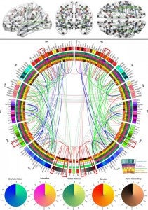
EMBARGOED FOR RELEASE 12 A.M. PT/3 A.M. ET, February 11, 2014 — For the first time, neuroscientists have systematically identified the white matter “scaffold” of the human brain, the critical communications network that supports brain function.
Their work, published Feb. 11 in the open-source journal Frontiers in Human Neuroscience, has major implications for understanding brain injury and disease. By detailing the connections that have the greatest influence over all other connections, the researchers offer not only a landmark first map of core white matter pathways, but also show which connections may be most vulnerable to damage.
“We coined the term white matter ‘scaffold’ because this network defines the information architecture which supports brain function,” said senior author John Darrell Van Horn of the USC Institute for Neuroimaging and Informatics and the Laboratory of Neuro Imaging at USC.
“While all connections in the brain have their importance, there are particular links which are the major players,” Van Horn said.
Using MRI data from a large sample of 110 individuals, lead author Andrei Irimia, also of the USC Institute for Neuroimaging and Informatics, and Van Horn systematically simulated the effects of damaging each white matter pathway.
They found that the most important areas of white and gray matter don’t always overlap. Gray matter is the outermost portion of the brain containing the neurons where information is processed and stored. Past research has identified the areas of gray matter that are disproportionately affected by injury.
But the current study shows that the most vulnerable white matter pathways — the core “scaffolding” — are not necessarily just the connections among the most vulnerable areas of gray matter, helping explain why seemingly small brain injuries may have such devastating effects.
“Sometimes people experience a head injury which seems severe but from which they are able to recover. On the other hand, some people have a seemingly small injury which has very serious clinical effects,” says Van Horn, associate professor of neurology at the Keck School of Medicine of USC. “This research helps us to better address clinical challenges such as traumatic brain injury and to determine what makes certain white matter pathways particularly vulnerable and important.”
The researchers compare their brain imaging analysis to models used for understanding social networks. To get a sense of how the brain works, Irimia and Van Horn did not focus only on the most prominent gray matter nodes — which are akin to the individuals within a social network. Nor did they merely look at how connected those nodes are.
Rather, they also examined the strength of these white matter connections, i.e. which connections seemed to be particularly sensitive or to cause the greatest repercussions across the network when removed. Those connections which created the greatest changes form the network “scaffold.”
“Just as when you remove the internet connection to your computer you won’t get your email anymore, there are white matter pathways which result in large scale communication failures in the brain when damaged,” Van Horn said.
When white matter pathways are damaged, brain areas served by those connections may wither or have their functions taken over by other brain regions, the researchers explain. Irimia and Van Horn’s research on core white matter connections is part of a worldwide scientific effort to map the 100 billion neurons and 1,000 trillion connections in the living human brain, led by the Human Connectome Project and the Laboratory of Neuro Imaging at USC.
Irimia notes that, “these new findings on the brain’s network scaffold help inform clinicians about the neurological impacts of brain diseases such as multiple sclerosis, Alzheimer’s disease, as well as major brain injury. Sports organizations, the military and the US government have considerable interest in understanding brain disorders, and our work contributes to that of other scientists in this exciting era for brain research.”
The research was supported by NIH grants 2U54EB005149-06, 1RC1MH088194, R41NS081792-01.
For a PDF of the study, press images or to arrange an interview with a researcher, contact Suzanne Wu at suzanne.wu@usc.edu.
About the USC Institute for Neuroimaging and Informatics
The Institute for Neuroimaging and Informatics aims to enhance discovery through the application of imaging and information technologies in the study of the brain. The Institute is dedicated to excellence in data acquisition, analysis, stewardship and computational innovation for the purpose of biomedical research.
About Digital USC
Digital USC is a university-wide initiative of $1 billion over 10 years towards gathering, interpreting and applying digital data on a massive scale. An international leader in fields from artificial intelligence to game design, with the largest computer science research program among American universities, USC is advancing knowledge in the development of virtual humans for therapy, entertainment, or historic preservation; the simulation and modeling of earthquakes and other complex systems; brain mapping; archiving, indexing and analysis of visual material; visual storytelling and data visualization; and in creating advanced online platforms for distance education and professional development.


