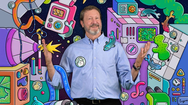
As USC’s director of science initiatives and director of the Translational Imaging Center, Scott Fraser has a hand in a wide array of research endeavors, from applied physics to neurobiology. (Illustration/Brosmind)
Scott Fraser is an explorer through invention
Scientific polymath Scott Fraser has a special talent for the live imaging of microscopic organisms — among a multitude of skills.
As a kid, Scott Fraser took his toys apart to see how they worked, and he delighted at the discovery of hidden mechanisms and unexpected connections. As a young man, he labored alongside his grandfather, a carpenter who built the forms for many of the poured-concrete bridges in Pasadena, and later applied his penchant for building things to science with his adviser, Daniel Petersen, at Harvey Mudd College.
Those early forays in engineering have turned Fraser into what he is today: a physicist, engineer and developmental biologist with a special talent for the live imaging of microscopic organisms. Delighting in the unexpected has helped him find surprising solutions to scientific problems: He’s learned the trick of using a human eyelash to cut through a frog embryo. In his lab, he turned an airport baggage scanner into a low-light microscope for a better view of tiny biological specimens.
As USC’s director of science initiatives and director of the Translational Imaging Center, Fraser has a hand in a wide array of research endeavors, from applied physics to neurobiology. And while his list of scientific publications, citations and patents keeps growing — he was named to the National Academy of Inventors in 2016 and the National Academy of Medicine in 2020 — his most influential contribution to science may be his work as a mentor and collaborator.
A force multiplier
Fraser, who recently marked a decade at USC after 21 years at Caltech, is known for solving other people’s problems and teaching others to do the same. One of Fraser’s favorite innovations, still central to his lab today, is a gadget for encouraging scientific discovery:
A restaurant-grade espresso machine.
“Everybody and their dog started coming to my conference room to make a coffee and sit down,” Fraser recalls. ”They came after something worked or after something failed. They would be elated or discouraged, but they would talk to the other people there grabbing coffee. So, in the first year of that coffee machine, there must have been two dozen patents filed.”
More than 120 trainees have passed through his labs at USC and Caltech. Nine are current USC scientists. Others landed at Harvard and Stanford universities, Children’s Hospital LA and dozens of other institutions and companies.
“In many labs, when people leave, they go from being a family member to being a competitor — the ‘enemy.’ We’ve tried to minimize that by encouraging scientists when they leave to take their project with them, if they want to,” Fraser says. “Afterwards, it’s sort of the empty nest syndrome. You think, ‘What am I going to do now?’ It forces the lab to reinvent itself.”
Andrea Armani, who holds the Ray Irani Chair in Chemical Engineering and Materials Science at USC, was a postdoc in Fraser’s lab from 2006 to 2008. She pursued — and was paid for — her interests in integrated biosensors, among other things, even though the Fraser Lab had other projects in the works.
“Usually, faculty members want to hire postdocs who they can plop right in (to ongoing projects), and they’re going to be immediate contributors,” Armani says. “I came in, and I was not an immediate contributor. But I learned an immense amount. He was always incredibly supportive of my training and that was invaluable.”
Innovation at the smallest scale
Like the late oceanic scientist Jacques Piccard, Fraser is an explorer through invention. While Piccard designed and built deep-sea ships to explore distant realms in the ocean, Fraser designs and builds microscopes and other tools to expand what we can see. He calls it “the world’s smallest reality TV show studio.”
For example, Fraser, with Don Arnold, Carl Kesselman and other collaborators at USC, used a custom-made microscope and fluorescent markers to film a live zebrafish forming a memory in real time. Zebrafish are a popular model for research because of their fully sequenced genome, rapid development, easy transgenesis with fluorescent proteins, and their nearly transparent embryo. Findings in zebrafish frequently yield insights about humans.
The study was groundbreaking, revealing for the first time, through images and movies, how memories are formed. It opened up new possibilities for understanding brain development or the destruction of memory by Alzheimer’s or other age-related diseases.
If a picture is worth a thousand words, “a movie is worth millions of words,” Fraser says.
An idea born over beers
It is one thing to ponder the secrets of life; it’s another thing to film them. For a recent, Harvard-led paper published in Science, Fraser helped answer a long-standing question in developmental biology: the difference between right and left. ?The study explains how zebrafish embryos “break symmetry” as their bodies take shape in their first 24 hours of life. Although zebrafish (and human) bodies are externally symmetric across the left-right axis, as an embryo grows there are important left-right asymmetries in the shape and positioning of most internal organs, reflected in the positioning of the heart, stomach and spleen.
Until now, the signals that kick off that process were a mystery.
“Left-right asymmetry is important for everything,” Fraser says. “For example, your heart gets placed to one side. People who have randomized asymmetry, or reversed asymmetry, end up having shorter lifespans. They sometimes die early or never make it through birth because their major blood vessels don’t hook up right.”
The idea for the experiment was born over beers nearly 10 years ago, after a class on imaging techniques that Fraser was teaching at the Woods Hole Marine Biological Laboratory. He invited a Yale postdoc, whom he’d just met, to visit the Translational Imaging Center at USC.
“One of the crazy ideas Scott and I came up with over beers was: Wow, wouldn’t it be cool to address this fundamental, mysterious question in our field using a pure laser approach to apply force onto cilia?” recalls Shiaulou Yuan, who was a postdoc at Yale. “About six months later, I went to the USC Translational Imaging Center, and they built the first prototype instrument. Really incredible. I didn’t know Scott at all before this. I just showed up.”
During weeklong visits, Yuan and Thai Truong, then a postdoc in Fraser’s lab, put in long days for the next few years. They’d start in the morning when the zebrafish eggs were laid and conduct experiments around midnight, when the embryos had matured enough to have a cilia-lined “left-right organizer,” a temporary orb responsible for asymmetrical growth.
Truong, now an assistant professor (research) who describes himself as Fraser’s chief “MacGyver,” combined optical “tweezers” — tiny lasers used for positioning microscopic materials — with another imaging technique called light sheet microscopy. The tools allowed them to see how a clockwise swirl of liquid inside the orb triggered cilia to send a chemical message. They solved the puzzle: Movement of the liquid, not some signaling molecule, provided the signal.
“I can’t think of any other lab in the world that would have the interest in doing this without any gain. It’s unclear what [Fraser] would gain out of this,” says Yuan, now an assistant professor of medicine at Massachusetts General Hospital and Harvard Medical School. “He’s a wonderful mentor to young scientists like myself. I wasn’t in his lab and we didn’t know each other, but he enabled me to do this fantasy-type experiment. He helped me establish my career, my lab here in Boston.”
Seeing a frog embryo for the first time
For Fraser, these mysteries are what drew him to embryology in the first place. As an undergraduate, he had been a physics student with a specialty in developing low-noise electronics and deploying vibration control. He thought optics would be a tool, not a huge part of his future. But then, as a grad student, he made the “mistake” of looking through a friend’s microscope and seeing a frog embryo.
“It was a moment — I guess you call it an epiphany — but it was phenomenal to see the cells develop, to watch how after a microsurgery, the cells could heal so quickly.”
Fraser had so many questions. And the answer to many of them was, “We don’t know.”
“And that,” he says, “seemed fantastic.”


